Flow Acceleration Towards and Into a Long Segmental Stenosis:
Doppler Ultrasound
by | AHA 1992 | Publications, Doppler Ultrasound
Flow Acceleration Towards and Into a Long Segmental Stenosis: Spatial Movement of the Vena Contracta: An In Vitro Study Using Digital Color Doppler
Takahiro Shiota, Dag Teien, You-Bin Deng, Sage Ewing, Robin Shandas, David J. Sahn Univ Calif, San Diego, CA
Presented by: Takahiro Shiota (David Sahn)
AHA 1992
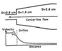
We used Color Doppler (CD) to estimate changes in flow velocity (VEL) when fluid accelerates towards and into a long segment stenosis in a modified pulsatile flow phantom (EchoCal CD 10) with calibrated VELs. Fluid enters the system through a 2.6 cm (diameter) cylinder to accelerate through a tapering segment ending in an abrupt orifice (OR) into a 0.8 cm diameter tube. We used a Ving-Med 750 scanner with on-line transfer of digital CD VEL data to a Macintosh IIci computer. Maximum VELs (MAX-VELs) ranging from 1.0 m/sec to 5.0 m/sec were recorded by CW Doppler in the narrowest section. Correct CD determination of VELs along the tapering segment was possible after alias unwrapping (r=0.98 to actual VEL). For MAXVEL of 1.0 m/sec, center-line VEL increased from 0.12 cm/sec at the inlet of the tube to 0.71 m/sec just proximal to the OR. MAXVEL was located 0.19 cm distal to the OR (vena contracta, VC) and within the tube. Similar findings of VC displacement were noted for other MAXVELs. The distance from the OR to VC correlated well with MAXVEL (r=0.96, p < 0.0001). In our study, digital CD analysis provided accurate spatial VEL profiles for accelerating flows and provided information about spatial displacement of the VC as a function of MAXVEL.


